Call Us Today!
(425) 999-8159
New Patients
(425) 264-7102


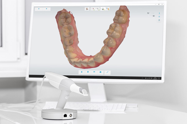
The iTero® Intraoral Scanner is a handheld digital imaging system that captures highly detailed three-dimensional images of teeth, gums, and bite relationships. Unlike traditional putty impressions, the scanner produces an instant, digital model that clinicians can manipulate and examine on-screen. This capability changes how dentists diagnose, plan, and communicate treatment by converting physical impressions into precise virtual data that can be used immediately.
At the office of 360 Dental of Mill Creek, we rely on this technology to speed up workflows and increase diagnostic confidence. The scanner’s high-resolution captures help reveal subtle surface details and occlusal contacts that might be difficult to appreciate with conventional methods. For clinicians, that means fewer surprises during restorative or orthodontic procedures and a clearer roadmap for treatment.
Beyond the immediate imaging advantages, digital scans provide a permanent, shareable record that integrates with many restorative and orthodontic systems. The iTero® platform supports alignment with lab software, milling centers, and aligner manufacturers, making it a central tool in modern dental care. This interoperability helps teams deliver consistent, predictable outcomes across a range of services.
A scanning appointment with the iTero® system begins with a quick orientation and a brief review of what will be captured. The clinician gently guides the scanner’s wand through the mouth, pausing at key zones such as the biting surfaces, interproximal contacts, and gum margins. The process is non-invasive and generally well tolerated by most patients because it removes the need for bulky impression trays and impression material.
The scanner’s guided workflow and real-time feedback allow clinicians to verify coverage as they go, so missing areas can be corrected immediately. Scans that once took multiple attempts with traditional materials can now be completed in a single visit. This reliability shortens chair time while preserving clinician oversight and control over the final digital model.
Once the capture is complete, the digital model is available within minutes for analysis, treatment simulation, or transfer to a lab partner. Patients can view the images on-screen with the clinician, enabling more meaningful conversations about treatment options. The transparency and immediacy of the digital scan enhance informed decision-making for both routine and complex cases.
Clinically, the iTero® Intraoral Scanner improves the fit and function of restorations by delivering accurate margins and occlusal relationships to dental laboratories and milling centers. For crowns, inlays, and onlays, this precision reduces the need for adjustments and remakes. The scanner’s fine detail helps technicians fabricate restorations that align closely with the patient’s natural anatomy, which supports longevity and comfort.
In orthodontics, the scanner provides exact digital impressions used to design clear aligners and other appliances. Because the digital scan captures the full three-dimensional relationship of the teeth, treatment simulations are more accurate and aligner staging can be better planned. This results in clearer treatment goals and helps clinicians set realistic expectations about timeline and outcomes.
For implant planning and complex restorative cases, the iTero® data can be combined with other digital records—such as CBCT scans—to create coordinated, multidisciplinary treatment plans. This integrated approach allows for more predictable surgical guides, prosthetic components, and final restorations, improving both functional results and patient satisfaction.
Digital impressions streamline communication between the dental office and external partners by providing instantly transferable files. When a scan is sent to a laboratory or specialist, there is no waiting for physical models to ship and no degradation of detail from impression material handling. This faster exchange reduces turnaround time while preserving image fidelity for fabrication or further planning.
Because the iTero® system aligns with many commonly used digital platforms, it supports a smoother handoff to CAD/CAM workflows and aligner manufacturers. Technicians receive standardized, high-quality data that simplifies the design process and minimizes back-and-forth adjustments. For the treating clinician, that reliability often translates to fewer in-office visits and more predictable final appointments.
In addition to external collaboration, in-house teams benefit from centralized digital records that integrate with practice management and imaging software. This consolidated information improves case tracking, quality control, and patient education, allowing the practice to maintain higher standards across restorative, orthodontic, and surgical services.
Patients frequently comment on the comfort of a digital scan compared with traditional impressions. The scanning wand is compact and operates without the gag-inducing materials used in some conventional impression techniques. This less intrusive experience is especially appreciated by those with sensitive gag reflexes, limited mouth opening, or dental anxiety.
The visual nature of the scan fosters clearer conversations about oral health and treatment choices. By viewing a 3D model alongside the clinician, patients can more readily understand problem areas, proposed corrections, and projected outcomes. This shared view supports collaborative decision-making and helps patients feel more confident about their care path.
Finally, the permanence and portability of digital records mean that future appointments can build on prior scans without repeating impressions unnecessarily. Whether tracking wear patterns, monitoring restorative margins, or evaluating orthodontic movement, the digital archive becomes a useful reference that benefits clinical continuity and patient convenience.
In summary, the iTero® Intraoral Scanner brings precision, comfort, and interoperability to modern dental care. By converting intraoral anatomy into accurate, shareable digital models, it supports better restorative fit, clearer orthodontic planning, and more efficient collaboration with labs and specialists. If you would like to learn more about how this technology is used in our practice, please contact 360 Dental of Mill Creek for additional information.
The iTero® intraoral scanner is a handheld optical device that captures a series of high-resolution images of the teeth, gums and bite relationships and converts them into a three-dimensional digital model. The clinician guides the wand through the mouth while the scanner’s software stitches the images together in real time to produce an accurate virtual representation of intraoral anatomy. This process replaces physical impression materials with an immediate, manipulable digital file that can be used for diagnosis and treatment planning.
The resulting digital model can be viewed from multiple angles and measured on-screen, which aids communication between clinician and patient as well as coordination with laboratories and specialists. Scans are available within minutes for analysis, simulation, or export to CAD/CAM and aligner manufacturers. Because the data is digital, it can be archived, compared over time, and integrated with other imaging modalities when needed.
Digital scans eliminate the need for impression trays and putty by creating a virtual model directly from intraoral captures, which reduces mess and the potential for distortions associated with material handling. Traditional impressions rely on the physical properties of impression materials and stone models, which can introduce dimensional changes during pouring and shipping. A digital workflow produces standardized files that preserve fine surface detail and occlusal relationships without the same susceptibility to material shrinkage or tear.
Because clinicians can verify scan coverage in real time, missing areas are identified and corrected immediately, often reducing the need for repeat captures. Digital files are also transferable instantly to labs and specialists, which shortens turnarounds compared with shipping physical models. The result is a more efficient, predictable handoff for restorative and orthodontic workflows.
A scanning visit typically begins with a brief orientation where the clinician explains the areas to be captured and what the images will show. The practitioner then moves the compact scanner wand around the dental arches, pausing at key zones like biting surfaces, gum margins, and interproximal contacts to ensure complete coverage. The process is noninvasive and generally completed in a single session without the use of impression material.
Patients can often watch the scan as it progresses on-screen, which helps illustrate findings and planned treatments. The clinician verifies coverage during the capture, so any gaps are corrected on the spot and the finalized model is available within minutes for review or export. This workflow tends to shorten chair time and enhance clarity about next steps.
The iTero® scanner delivers precise margin detail and occlusal relationships to dental laboratories and milling centers, which supports the fabrication of restorations that fit more accurately on delivery. Accurate digital impressions reduce the likelihood of ill-fitting crowns, inlays or onlays and can minimize the need for adjustments and remakes. Technicians use the fine detail captured by the scanner to reproduce natural anatomy and contacts that improve function and comfort.
Additionally, digital models can be integrated with in-office CAD/CAM systems or sent to external labs with consistent, standardized data, which streamlines production and quality control. The ability to compare scans over time also helps clinicians monitor restorative margins and adjacent tissue health to address issues proactively. Overall, the digital workflow contributes to more predictable restorative appointments and better long-term results.
For orthodontics, the iTero® scan provides an exact three-dimensional record of tooth positions that is used to plan and stage aligner therapy with greater accuracy. Treatment simulations based on digital scans allow clinicians and patients to visualize projected movements and set realistic expectations for timelines. Aligners and appliances manufactured from high-quality digital data typically require fewer adjustments during treatment because staging is planned from precise models.
Because scans are digital and interoperable with many aligner systems, files can be sent directly to aligner manufacturers for design and fabrication without converting physical impressions. This direct transfer reduces potential errors and preserves the fidelity of occlusal relationships and interproximal detail. The result is smoother case coordination and clearer communication about treatment goals.
Yes, iTero® digital scans can be combined with other diagnostic records, such as cone-beam CT data, to support coordinated implant planning and guide fabrication. Merging surface scans with volumetric imaging helps clinicians align prosthetic designs with underlying bone anatomy to plan implant position, angulation and emergence profiles more predictably. This multidisciplinary integration aids the design of surgical guides and provisional restorations that fit the planned restorative outcome.
When scans are exported in compatible formats, laboratories and guide manufacturers can use the unified dataset to produce components that match the planned prosthetic contours and implant positions. Having accurate digital models reduces intraoperative surprises and contributes to a more seamless transition from surgical placement to final restoration. The archival nature of digital records also supports long-term follow-up and maintenance.
The iTero® platform generates standardized digital files that can be transferred instantly to external laboratories and specialist partners, removing delays associated with shipping physical impressions and models. Digital transfers preserve surface detail and occlusal relationships without the risk of damage or distortion that can occur during handling. Labs receive consistent, high-quality data that simplifies design and fabrication workflows.
Instant file sharing reduces turnaround time and minimizes back-and-forth communications by providing technicians with the information they need up front. This streamlined exchange often leads to fewer in-office adjustments and more predictable appointment scheduling for final delivery. Centralized digital records also make it easier for specialists to review cases and provide coordinated input when multidisciplinary planning is required.
Patients commonly report that digital scanning is more comfortable than conventional impressions because it avoids bulky trays and impression materials that can trigger gagging or discomfort. The scanner wand is compact and designed for gentle intraoral movement, which makes it suitable for patients with limited mouth opening or sensitive reflexes. The procedure is noninvasive and does not expose patients to ionizing radiation since it captures surface images only.
Clinicians can pause and address sensitivity or adjust technique during the capture to maximize patient comfort, and real-time visualization helps reassure patients about what is being recorded. Hygiene protocols for scanner sleeves and infection control are followed in the same way as other intraoral devices. Overall, scanning provides a safe, patient-friendly alternative to traditional impression techniques.
Digital scans are archived as part of the patient’s electronic record and can be retrieved for comparison, treatment planning or remakes without requiring new impressions in many cases. This longitudinal archive supports monitoring of wear patterns, restorative margins and orthodontic movement over time, enabling clinicians to detect changes and intervene early when necessary. Because scans are portable files, they can also be shared with specialists or transferred to new systems when clinically appropriate.
Integration with practice management and imaging software helps maintain case histories and ensures that digital models remain accessible for future restorative, orthodontic or surgical needs. Storing scans digitally reduces the need for physical model storage and preserves detail indefinitely when maintained with appropriate backup and security measures. Patients benefit from continuity of care because prior scans inform subsequent treatment decisions.
The iTero® scanner supports the practice’s commitment to precise, predictable dentistry by providing accurate digital models that enhance diagnosis, treatment planning and laboratory communication. Using a modern digital workflow helps the clinical team reduce chair time, verify capture quality in real time and coordinate care with specialists and labs more efficiently. These capabilities align with the practice’s focus on delivering personalized, results-driven treatments.
Digital scans also improve patient understanding by allowing clinicians to review three-dimensional images on-screen and discuss treatment options with visual clarity. The archived digital records support continuity of care for restorative, orthodontic and surgical services and provide a reliable reference for monitoring changes over time. Employing digital impression technology is one way the office maintains contemporary standards of care for patients in Mill Creek and the surrounding community.