Call Us Today!
(425) 999-8159
New Patients
(425) 264-7102


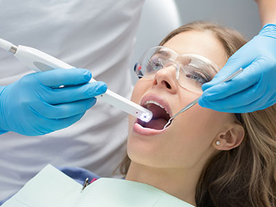
An intraoral camera is a compact, pen-sized imaging tool designed to capture detailed, full-color pictures inside the mouth. Equipped with a small lens and high-resolution sensor, it provides clear visual access to tooth surfaces, gum tissue, and hard-to-see areas that are difficult to inspect with the naked eye alone. Images appear in real time on a monitor, giving clinicians an immediate, magnified view of the oral cavity.
Because the device is portable and maneuverable, it can be positioned to examine individual teeth, restoration margins, and areas beneath orthodontic appliances. The clarity of the images helps the dental team identify tiny fractures, early decay, worn restorations, and inflammation—issues that may not be obvious during a routine visual exam. Intraoral cameras are a noninvasive complement to tactile examinations and radiographs.
Beyond simple photography, modern intraoral cameras often include features such as LED lighting, autofocus, and the ability to capture stills and short video clips. These capabilities turn a routine checkup into a more thorough diagnostic process, allowing clinicians to document findings and track changes over time with a level of precision that supports better clinical decisions.
Intraoral images give clinicians a magnified, illuminated view that improves detection of subtle issues. Small cracks in enamel, margin gaps around crowns and fillings, and early carious lesions can be more readily identified when viewed at higher magnification. This detailed visualization reduces reliance on guesswork and helps prioritize which areas need further investigation or immediate treatment.
When combined with other diagnostic tools—such as radiographs and periodontal measurements—intraoral images create a fuller clinical picture. The camera can document the progression of lesions or the condition of restorative work, which aids in deciding on conservative versus restorative approaches. Accurate imaging also helps in planning minimally invasive care by pinpointing the exact location and extent of a problem.
Because these images are saved to a patient’s record, they provide a consistent reference point for follow-up visits. Clinicians can compare sequential images to see how a condition responds to treatment or to identify trends that might otherwise be missed. This objective visual history strengthens clinical assessments and leads to more predictable treatment outcomes.
Seeing a close-up image of their own mouth often changes how patients perceive their oral health. Intraoral photography brings transparency to the exam process by letting patients view conditions in real time while the dentist explains what the images reveal. This direct visual communication helps patients make informed decisions about recommended care without needing extensive technical explanation.
For many patients, the ability to view magnified images makes it easier to understand why a particular area needs attention. Visual evidence clarifies abstract descriptions—such as “a small fracture” or “early decay”—and helps align patient expectations with the clinician’s recommendations. It also empowers patients to monitor progress after treatment and reinforces the importance of daily preventive habits.
Because the camera is gentle and quick to use, the patient experience remains comfortable. Captured images can be shown immediately, annotated or magnified during the visit, and placed into the patient’s file for future reference. That clarity fosters collaboration between patient and provider and supports shared decision-making about care goals.
Intraoral cameras integrate seamlessly with modern dental record systems, allowing images to be stored alongside x-rays, clinical notes, and treatment plans. This digital integration streamlines documentation, simplifies coordination with specialists or laboratories, and supports efficient case presentation. When a clinician needs to communicate specifics—such as restoration margins or an area of concern—high-quality images convey details more effectively than text alone.
Images captured by the intraoral camera are also useful when collaborating with dental laboratories for crowns, veneers, or other prosthetics. Clear photos of occlusal surfaces and preparation margins provide the lab with valuable visual context that can improve the fit and aesthetics of final restorations. Similarly, visual records facilitate professional referrals by giving colleagues a precise view of the issue before a transfer of care occurs.
Incorporating intraoral photos into the treatment workflow enhances efficiency: clinicians can document findings quickly, update records in real time, and present treatment options in a visual format that patients readily understand. The result is a smoother clinical process with fewer surprises and better alignment between planned and delivered care.
Intraoral cameras are designed with patient safety and infection control in mind. Clinicians use protective sleeves or barriers for each patient and follow established sterilization protocols for any reusable components. These standard precautions ensure the device can be used repeatedly in a busy practice without compromising hygiene or safety.
On the information side, images captured by the camera are managed within secure, electronic health record systems. Proper handling means images are stored in a way that respects patient privacy and regulatory requirements, and access is limited to authorized members of the dental team. Secure image management supports continuity of care while protecting sensitive health information.
Routine maintenance and periodic software updates keep camera performance reliable and image quality consistent. Staff training on proper handling, image capture techniques, and data management ensures that the diagnostic value of intraoral photography is fully realized in everyday practice while maintaining patient comfort and confidentiality.
At the office of 360 Dental of Mill Creek, intraoral cameras are one of several tools we use to provide clear, patient-centered care. If you’d like to learn more about how this technology is used during an exam or how images are handled in your chart, please contact us for more information.
An intraoral camera is a small, handheld imaging device used to capture high-resolution, full-color pictures of the inside of the mouth. The unit typically includes a tiny lens, LED lighting, and a sensor that transmits images in real time to a monitor for immediate viewing. Because it produces magnified views, the camera allows clinicians to inspect tooth surfaces, gum tissue, and other areas that are difficult to see with the naked eye.
Modern intraoral cameras can take still photos and short video clips, and many feature autofocus and image-enhancement tools. These capabilities help clinicians document findings, track changes over time, and communicate conditions clearly to patients. Used alongside other diagnostic methods, the camera improves the thoroughness of routine exams without increasing invasiveness.
An intraoral camera provides a magnified, illuminated view that makes it easier to detect subtle problems such as hairline cracks, small areas of decay, and margin gaps around restorations. This enhanced visualization reduces reliance on guesswork and helps clinicians prioritize which areas require further testing or early intervention. Clear images can reveal surface textures, color changes, and anatomical details that might otherwise be overlooked.
When combined with radiographs, periodontal measurements, and clinical probing, intraoral images contribute to a more complete diagnostic picture. The ability to save sequential images to the chart also supports monitoring of lesion progression or healing after treatment. Overall, the technology supports more accurate, conservative, and predictable clinical decision-making.
During an exam, you will often see live, magnified images of your teeth and gums displayed on a chairside monitor as the clinician guides the camera. The dentist or hygienist may pause to capture key photos, point out areas of concern, and annotate images to illustrate what they are describing. This visual approach helps you understand the exact location and nature of an issue without relying solely on verbal explanation.
Our team at the office of 360 Dental of Mill Creek uses these images to explain treatment options and expected outcomes in a clear, evidence-based way. Viewing your own images can make it easier to weigh preventive measures versus restorative care and to follow recommended home care steps. The process is quick and designed to enhance communication between you and your provider.
Imaging with an intraoral camera is noninvasive and safe when used according to standard infection‑control protocols. Clinicians place single‑use protective sleeves or barriers over the camera for each patient and follow sterilization procedures for any reusable components. These precautions prevent cross-contamination while allowing the device to be used repeatedly during a busy clinic day.
In addition to physical barriers, routine maintenance and periodic software updates help ensure consistent image quality and reliable performance. Staff training on proper handling and capture techniques further protects patient comfort and the diagnostic value of the images. Taken together, these measures make intraoral imaging both hygienic and patient-friendly.
Intraoral images are typically integrated into the patient’s electronic health record and stored on secure systems that protect privacy and limit access to authorized personnel. Images are labeled and archived with the date and clinical notes so they can be referenced during future visits or included in treatment documentation. Proper data management practices ensure images remain part of a patient’s legal and clinical record.
When collaboration is needed, clinicians can share selected images with specialists or dental laboratories as part of a referral or case planning process, usually with the patient’s consent. Secure transfer methods are used to protect sensitive health information while providing colleagues with the visual detail necessary for coordinated care. Patients may also request copies of images through the practice’s standard record‑release procedures.
No, an intraoral camera does not replace dental X-rays; rather, it complements radiographic imaging by providing detailed surface views that X-rays cannot show. Cameras are excellent for identifying visible surface problems, recording the condition of restorations, and educating patients about what the clinician sees. X-rays, in contrast, reveal internal structures such as tooth roots, bone levels, and interproximal decay beneath contact points.
For comprehensive diagnosis, dentists use both tools together so each method’s strengths offset the other’s limitations. In many cases, a combined approach leads to earlier detection of disease and more targeted, minimally invasive treatment plans. A clinician will select the appropriate mix of visual, tactile, and radiographic diagnostics based on each patient’s needs.
Intraoral images help clinicians document restoration margins, preparation details, and occlusal relationships that are important for crowns, veneers, and other prosthetics. High-quality photos can be sent to dental laboratories to give technicians a precise visual reference for shade, contour, and margin placement, which improves the accuracy and aesthetics of final restorations. Visual records also allow the dental team to compare preoperative and postoperative outcomes objectively.
For cosmetic planning, intraoral photography enables side‑by‑side comparisons and visual simulations that clarify expectations and goals. Seeing a visual record lets patients participate in shared decision‑making and helps clinicians design conservative approaches that preserve healthy tooth structure. The result is more predictable restorative work and higher alignment between planned and delivered results.
An intraoral camera captures two‑dimensional still images or video of the mouth’s surfaces, while an intraoral scanner records three‑dimensional digital impressions that can be used to design restorations and appliances. Cameras are optimized for visualization and documentation, making them ideal for patient education and surface assessment. Scanners, on the other hand, generate precise 3D models used in CAD/CAM workflows and laboratory fabrication.
Both technologies are valuable in modern dentistry and often serve complementary roles: the camera for diagnostic clarity and communication, and the scanner for digital impressioning and production. A clinician will choose the tool that best fits the diagnostic goal or treatment workflow for a particular case. Understanding the strengths of each device helps set appropriate expectations for how they contribute to care.
Yes, intraoral cameras are limited to surface visualization and cannot provide information about structures beneath the enamel or inside the jawbone. They are not effective at detecting deep interproximal decay, root infections, or bone loss on their own, which is why clinicians rely on radiographs and clinical tests as well. Camera images also depend on access and patient cooperation; tight contact areas or subgingival problems can be difficult to capture clearly.
Because of these constraints, cameras are used as one part of a multi‑modal diagnostic approach rather than a standalone solution. Clinicians interpret camera images alongside X-rays, periodontal charts, and tactile examination to form a complete diagnosis. When additional detail is needed, targeted imaging or specialist referral will be recommended to clarify the condition and guide treatment.
Capturing intraoral images is typically quick and efficient, often adding only a few minutes to a routine exam when multiple photos are taken. The camera is guided around the mouth to document specific areas, and clinicians select the most useful stills or short clips for the chart. Because the process is noninvasive and chairside, it fits smoothly into the clinical workflow and rarely causes discomfort.
At the office of 360 Dental of Mill Creek, clinicians can include intraoral images in your chart and make them available through standard record‑release or patient‑portal procedures. Patients who wish to review their images at home can request copies in an appropriate electronic format or receive printed summaries as part of their visit documentation. Providing images for review supports informed decision‑making and helps patients follow recommended care instructions.