Call Us Today!
(425) 999-8159
New Patients
(425) 264-7102


At the office of 360 Dental of Mill Creek, we use advanced imaging to make diagnoses clearer and treatments more predictable. Cone-beam computed tomography (CBCT) gives our team three-dimensional views of the mouth and facial structures that traditional two-dimensional X-rays cannot provide. These detailed images help us see anatomy with precision so we can plan treatments that prioritize long-term health and function.
CBCT is not a single solution for every situation, but it is an indispensable tool when complex anatomy or surgical planning is involved. It complements the clinical exam and other digital records, giving clinicians the context they need to make informed decisions. When used appropriately, CBCT enhances safety, reduces surprises during treatment, and leads to better outcomes for patients.
Cone-beam CT uses a cone-shaped X-ray beam and a specialized detector to capture volumetric data in a single rotation. The result is a high-resolution 3D image—often called a volumetric dataset—that can be viewed in slices or reconstructed into lifelike models. This level of detail reveals spatial relationships among teeth, bone, nerves, and sinus cavities that planar radiographs may obscure.
Unlike conventional medical CT scanners, dental CBCT units are designed for focused imaging of the head and neck, which allows for lower radiation exposure and targeted scans. Clinicians can select the field of view to capture only the area of interest, minimizing unnecessary imaging while still obtaining diagnostically relevant information.
Because the images are digital and easily integrated into treatment software, they can be manipulated, measured, and shared with specialists or dental laboratories. This interoperability supports collaborative planning and ensures that everyone involved in a patient’s care is working from the same precise information.
One of the most common uses for CBCT in dental practices is implant treatment planning. Accurate assessment of bone volume, density, and the location of critical structures—such as the inferior alveolar nerve or the maxillary sinus—helps our team determine optimal implant size, orientation, and position. This reduces the risk of complications and supports predictable, long-term results.
CBCT also enables virtual surgery and the creation of surgical guides. With digital planning tools, clinicians can simulate implant placement in three dimensions and transfer that planned position to a guide used during the actual procedure. That workflow bridges the gap between planning and execution, improving both accuracy and efficiency.
Beyond implants, CBCT assists with many oral surgical procedures. It clarifies the path and depth of impacted teeth, identifies anatomical variations, and helps anticipate potential challenges—giving surgeons the information they need to proceed with confidence and care.
In cases where symptoms or conventional images leave questions unanswered, CBCT can provide the missing detail. For endodontic assessments, for example, CBCT can reveal root canal anatomy, hidden canals, or subtle fractures that are difficult to detect on traditional films. This allows for more targeted and effective treatment planning.
CBCT is also valuable for evaluating suspected pathology, traumatic injuries, and developmental anomalies. Its ability to display bone and soft-tissue relationships in three dimensions makes it easier to identify the extent and exact location of a problem, which is essential for accurate diagnosis and timely intervention.
Additionally, CBCT can play a role in airway and TMJ evaluations, giving clinicians a clearer picture of structural contributors to breathing or joint concerns. While not every airway or TMJ issue requires CBCT, when anatomical detail matters, the technology offers critical insight.
Modern CBCT scanners are designed with patient comfort and workflow efficiency in mind. Scans are completed quickly—often in less than a minute—and are performed with the patient seated or standing. The brief scan time helps reduce motion artifacts and makes the experience straightforward for most people, including those who may find longer procedures difficult.
Radiation exposure with dental CBCT is generally lower than medical CT and can be comparable to or modestly higher than a full-mouth series of intraoral X-rays, depending on the field of view and settings used. Our team follows the principle of ALARA (as low as reasonably achievable), tailoring each scan to capture only what is necessary for diagnosis or treatment planning.
Because CBCT data are captured digitally, images are available immediately for review and discussion. This allows clinicians to explain findings to patients in a visual and accessible way, improving understanding and helping patients participate in informed decisions about their care.
CBCT images are a natural fit for digital workflows. Scans can be combined with intraoral optical impressions and restorative design software to create comprehensive treatment plans—from implant prosthetics to full-arch restorations. This integration can streamline communication with dental laboratories and specialists, enabling coordinated care that aligns with the patient’s goals.
For orthodontic and restorative cases, three-dimensional data support better assessment of tooth position, root angulation, and spatial limitations. When combined with other digital records, CBCT contributes to more precise appliance design and placement, which can shorten treatment times and enhance final outcomes.
Furthermore, many clinicians use CBCT datasets to educate patients and to facilitate referrals when specialist input is needed. Clear visualizations of anatomy and planned interventions build trust and create a shared roadmap for treatment between providers and patients.
In summary, cone-beam computed tomography is a powerful diagnostic tool that enhances accuracy, improves treatment planning, and supports safer, more predictable dental care. When used thoughtfully as part of a comprehensive evaluation, CBCT gives clinicians the clarity they need to deliver high-quality results.
If you have questions about how CBCT might be applied to your care or would like to learn more about our imaging approach, please contact us for more information.
Cone-beam computed tomography, commonly called CBCT, is an advanced imaging modality that captures three-dimensional views of the teeth, jaws and surrounding facial structures. Unlike traditional two-dimensional X-rays, CBCT produces a volumetric dataset that can be viewed slice by slice or reconstructed into 3D models for detailed assessment. This additional spatial information reveals relationships among teeth, bone, nerves and sinuses that planar images may obscure.
CBCT units used in dentistry are specifically designed for focused imaging of the head and neck, allowing clinicians to select the field of view and limit exposure to the area of interest. The images are digital and interoperable with treatment planning software, which supports measurement, simulation and collaboration among providers. Because of its targeted capabilities, CBCT is used when three-dimensional detail materially affects diagnosis or treatment planning.
CBCT is recommended when clinical findings or conventional images do not provide sufficient information to make a confident diagnosis or plan treatment safely. Common indications include implant planning, evaluation of impacted or unusually positioned teeth, assessment of complex endodontic anatomy, and investigation of suspected pathology or facial trauma. It is also used selectively for TMJ assessment, airway considerations and cases with developmental anomalies where three-dimensional anatomy influences management.
Clinicians balance the diagnostic benefit of CBCT against the patient's needs and apply it selectively rather than routinely for all patients. The decision to image with CBCT follows a focused clinical exam and review of other digital records so that only the necessary area is scanned. This approach helps ensure imaging is targeted and clinically justified.
For implant dentistry, CBCT provides precise information about bone volume, bone quality and the position of critical anatomical structures such as the inferior alveolar nerve and maxillary sinuses. With accurate measurements in three dimensions, clinicians can determine optimal implant size, angulation and placement to achieve stable, functional restorations. This minimizes the risk of intraoperative surprises and supports predictable long-term outcomes.
CBCT datasets can be integrated with virtual planning tools to simulate implant placement and design surgical guides that transfer the planned position to the operative site. That digital workflow bridges planning and execution, improving both accuracy and efficiency during surgery. When combined with intraoral scans and restorative planning, CBCT helps align prosthetic goals with surgical execution for comprehensive implant treatment.
Dental CBCT scanners are engineered for focused imaging of the head and neck and typically deliver lower radiation doses than conventional medical CT scans. Exposure varies depending on the field of view, resolution settings and the specific unit, so clinicians select the smallest FOV and appropriate settings needed to address the clinical question. The practice of ALARA—keeping exposure as low as reasonably achievable—is followed to tailor each scan to the diagnostic need.
Before recommending CBCT, providers evaluate whether the expected diagnostic benefit outweighs the exposure and consider alternative imaging if appropriate. Protective measures, careful protocol selection and modern equipment help reduce unnecessary dose while preserving image quality. Patients with concerns about radiation should discuss risks and benefits with their clinician to make an informed decision.
Pregnancy and certain medical conditions may affect the appropriateness of imaging, so it is important to inform the dental team of relevant health information prior to scanning. In many cases, imaging can be postponed or alternative approaches used when pregnancy is confirmed. Clear communication allows the care team to choose options that prioritize patient safety and diagnostic value.
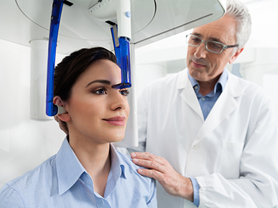
A CBCT scan is a brief, noninvasive procedure that is typically completed in under a minute with the patient seated or standing in the scanner. The patient is asked to remain still and may be positioned with a bite block or other support to stabilize the head during the rotation, which minimizes motion artifacts and improves image clarity. Metal objects such as jewelry or removable dental appliances are removed or adjusted to reduce interference in the images.
There is no direct contact with the scanner during the exposure, and the experience is generally comfortable for most patients, including those who find longer procedures difficult. Once the scan is complete the images are reconstructed quickly and become available for review, allowing the clinician to explain findings with visual aids. The concise nature of the exam supports efficient workflow and prompt discussion of next steps.
CBCT images are digital and can be exported, measured and integrated with treatment planning software, intraoral scans and laboratory workflows to create coordinated care plans. Specialists such as oral surgeons, endodontists and orthodontists can review the same volumetric dataset to assess anatomy, plan procedures and communicate recommendations with precision. This interoperability supports collaborative decision-making and reduces the risk of miscommunication about anatomical details.
Practically, CBCT data enable virtual surgery, guided implant fabrication, and enhanced endodontic assessment by revealing canal anatomy and pathology in three dimensions. The dataset also serves as a visual tool for patient education, helping clinicians illustrate findings and treatment options. By working from the same high-resolution information, care teams can align goals and streamline complex treatments.
Yes. The three-dimensional perspective of CBCT often reveals details that planar radiographs can obscure, such as subtle root fractures, accessory canals, extent of periapical pathology and the true relationship of lesions to adjacent anatomic structures. For endodontic cases, CBCT can identify additional canals, resorptive defects or apical pathology that may not be visible on conventional films. In trauma cases, CBCT provides accurate assessment of fracture lines and displacement.
CBCT is also valuable for evaluating the extent and location of cysts, tumors or inflammatory lesions within the jaws, which helps guide biopsy or surgical planning when needed. However, CBCT may not replace other imaging modalities when soft-tissue contrast is required; MRI or medical CT could be more appropriate for certain soft-tissue or complex maxillofacial concerns. Clinicians choose the imaging modality that best answers the clinical question.
Although CBCT is powerful for many dental applications, it is not indicated for every patient or clinical question and has limitations in soft-tissue contrast compared with MRI. Small fields of view are excellent for localized dental concerns but will not capture broader craniofacial anatomy, while larger fields increase exposure and may be unnecessary for routine care. Motion artifacts, metal scatter and patient cooperation can also affect image quality and interpretation.
Contraindications or cautions include confirmed pregnancy and situations where alternative, non-ionizing imaging would better address the diagnostic need. For very young patients, clinicians weigh the need for three-dimensional imaging against the ability to remain still and the cumulative exposure. Ultimately, use of CBCT is guided by clinical judgment, diagnostic necessity and patient-specific factors.
CBCT integrates seamlessly with intraoral scans, CAD/CAM design software and laboratory processes to support comprehensive digital workflows for restorative and surgical treatment. Combining volumetric data with optical impressions enables designers to plan implant-supported prostheses, design custom abutments and verify occlusion and emergence profiles before fabrication. This coordination helps align prosthetic outcomes with surgical planning and can reduce guesswork during restoration placement.
In orthodontics and full-arch rehabilitation, three-dimensional datasets support assessment of root positions, bone limitations and spatial relationships that inform appliance design and treatment sequencing. Shared digital records also simplify communication with external laboratories and specialists, allowing teams to work from the same precise datasets. The result is more predictable planning and streamlined delivery of complex care.
Your clinician will determine the appropriateness of CBCT after a thorough clinical examination and review of any existing imaging or records. If three-dimensional information is likely to change diagnosis or treatment planning, the team will explain the reasons for imaging, describe what to expect during the scan and discuss alternatives when applicable. This consultative approach ensures imaging is used thoughtfully and only when the expected diagnostic benefit justifies it.
If you have specific concerns about surgical planning, persistent symptoms, trauma or suspected pathology, mention these during your appointment so the clinician can assess whether CBCT will provide meaningful insight. The practice will also review safety considerations, address questions about the procedure and obtain consent before proceeding with any imaging. Clear communication helps ensure that care decisions are aligned with your needs and treatment goals.